
Benchtop SEM — Model: JCM-7000 NeoScope™
The JCM-7000 Benchtop Scanning Electron Microscope is designed based on a key concept of "Easy-to-use SEM with seamless navigation and live analysis".
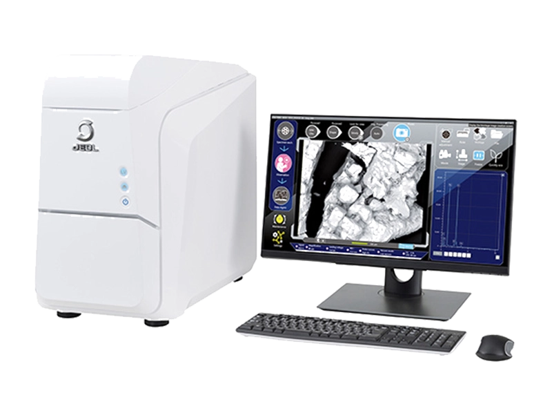
Manufacturer: JEOL — Japan
Overview
The JCM-7000 NeoScope™ benchtop scanning electron microscope closes the gap between the optical microscope and the SEM. In conventional SEM work, imaging and elemental analysis are separate steps — on the JCM-7000 they flow together: find the target on the optical image, zoom straight into the SEM image, and see the constituent elements displayed live during observation.
Features
Zeromag
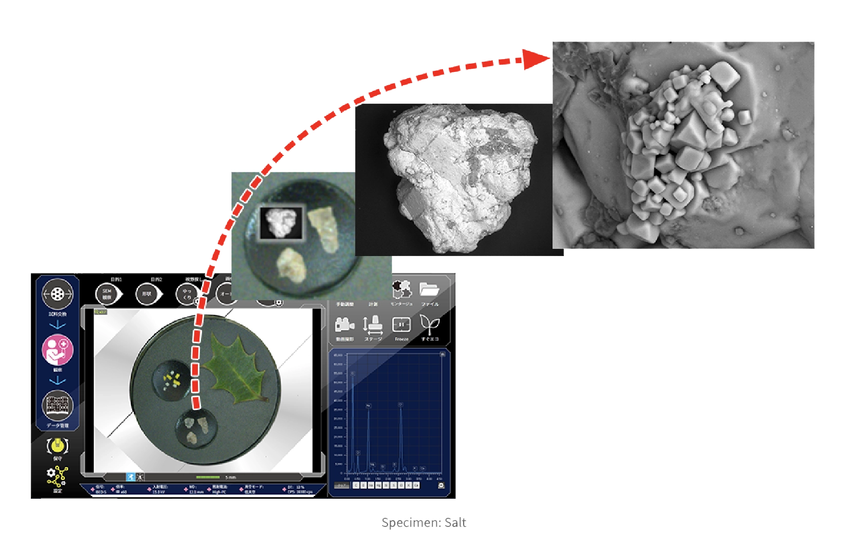
An optical image is acquired automatically when the sample is inserted. Search on the optical image and zoom in on the target — the view switches to the SEM image automatically, reaching the observation position in a minimal number of steps.
Live Analysis & Live Map
With Live Analysis, SEM observation and EDS analysis are no longer separate steps: the X-ray spectrum with the main constituent elements is displayed in real time on the observation screen. Live Map adds the spatial distribution of elements in real time, raising the odds of finding the elements of interest — and of catching unexpected ones. For detailed work, qualitative/quantitative analysis and element mapping are selected with the Purpose button and positioned directly on the observation screen. (An EDS system, option, is required.).
Auto Functions
Advanced auto functions provide clear images from low to high magnification.
Low Vacuum (LV) Mode
A 2-stage low-vacuum mode views non-conductive specimens — granules, leaves, rock salt — without any pre-treatment or conductive coating.
High Vacuum (HV) Mode
High-vacuum mode for detailed surface morphology.
Live 3D
The high-sensitivity 4-segmented backscattered electron detector acquires four SEM (BSE) images at once and displays a live 3D image — instantaneous shape assessment for complex topographies, with depth information included. Adding the optional SMILE VIEW™ Map software extends this to detailed 3D analysis such as surface-roughness measurement.
SMILE VIEW™ Lab
Links optical and SEM images, EDS data and locations for data review and reporting.
Improved Work Efficiency
Quickly confirm the morphology and composition that an optical microscope cannot identify.
Foreign-material analysis
Constituent elements are verified and foreign materials identified instantly — for example a black substance on a food product: locate it on the Zeromag optical image, double-click to centre it, zoom until the SEM image overlays automatically, and read the main constituent elements on screen. Report creation is easy, so feedback to the manufacturing site is fast.
Quality control
SEM reveals compositional contrast invisible to optical microscopy: the distribution of a white lubricant on white pharmaceutical granules shows clearly in the backscattered-electron compositional image, with no specimen pre-treatment in low-vacuum mode.
Screening before other instruments
Because observation and analysis need no pre-treatment, the same sample can go on to further analysis — thermal analysis, FT-IR, XRF and more — after the SEM measurement.
Easy Operation
Everyone should be able to use an SEM. The interface guides the workflow: buttons on the left lead the operation, buttons on top show the current procedure. Specimen exchange is a simple touch operation, and when a specimen is inserted an optical image of the entire 32 mm stage range is captured automatically (with the optional stage navigation system). Search the field of view on the optical image, click the target, press Auto — the SEM image is acquired. A motor-drive stage is standard, and data management is one button.
Easy maintenance
Changing the filament is easy: the electron gun uses a pre-centred cartridge integrated with the Wehnelt, replaced as a unit — fast exchange with correct filament positioning (the filament inside the cartridge is also replaceable). Alignment after replacement is fully automated.
No special utilities
The JCM-7000 operates from a 100 V service outlet; cooling water and liquid nitrogen are not required for SEM or EDS operation, and no special facilities are needed for installation.
One-click reporting
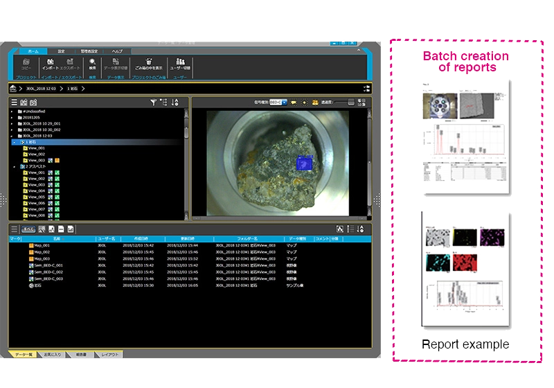
The Data management screen reviews or reanalyses data and generates batch reports covering everything from SEM images to analyses — exported to PDF, Microsoft Word or PowerPoint® with a single click.
Options to Extend Capabilities
Tilting & rotating motor drive holder
Observe specimens at various angles (0° to 45° shown); combined with the 2-axis motorized stage it provides 4-axis motorized control.
SMILE VIEW™ Map 3D analysis software
Stereo-pair and four-image 3D reconstruction, coloration and image editing; saved layouts/workflows repeat the same operation on new data; supports surface-texture standards such as ISO 25178 (Sq, Ssk, Sku, Sp, Sv, Sz, Sa).
Typical Applications
Metals
Conductive metal specimens are observed uncoated: striations and dimples on a SUS304 fracture surface reveal whether failure was ductile or brittle, and inclusions or fracture-origin materials can be analysed elementally (EDS option).
Glass & brittle fractures
Where optical microscopy struggles with transparent glass or plastics, the SEM finds the fracture starting point and reveals the top-surface morphology in detail.
Printed circuit boards
Low-vacuum mode suits composite materials — SEM observation and analysis without conductive coating, with Live 3D showing a defect's surface reconstruction and vertical dimension simultaneously.
Fibers & textiles
Complex fiber structures that are difficult to coat are observed in low vacuum, including elemental identification of foreign material lodged in carpet.
Food
Low vacuum is effective for water- and fat-rich foods; the optional LV cooling holder preserves heat-sensitive structures — for example mapping mineral distribution in processed cheese, uncoated.
Specifications
| Specifications | JCM-7000 |
|---|---|
| Resolution | 8.0 nm (Secondary electron image) |
| Magnification | ×10 to ×100,000 |
| Mode | High-Vacuum mode: Secondary electron image, Backscattered electron image (composition, topographic and shadow, 3D images) Low-Vacuum mode: Backscattered electron image (composition, topographic and shadow, 3D images) |
| Landing voltage | 5 kV, 10 kV, 15 kV (3 stages) |
| Electron source | Tungsten filament / Wehnelt integrated grid |
| Specimen stage | X-Y motor drive stage X: 40 mm Y: 40 mm |
| Maximum specimen size | 80 mm diameter × 50 mm height |
| Specimen exchange | Draw-out mechanism |
| Energy dispersive X-ray spectrometer (EDS) (optional) | SD detector with detection surface area: 30 mm² can be installed |
| Automatic functions | Alignment, focus, stigmator, brightness/contrast |
| Major installable options | Stage Navigation System; Tilting and Rotating Motor Drive Holder: Tilt: −10 to +45°, Rotation: 360°; EDS (energy-dispersive X-ray spectrometer): SD detector with detection surface area: 30 mm²; Particle Analysis Software 3; 3D Analysis Software (SMILE VIEW™ Map) |