
SEM — Model: JSM-IT510
The JSM-IT510, the newly added Simple SEM function allows users to "leave the manual repetitive operation to it", required for SEM observation, making SEM observation more efficient and easier.
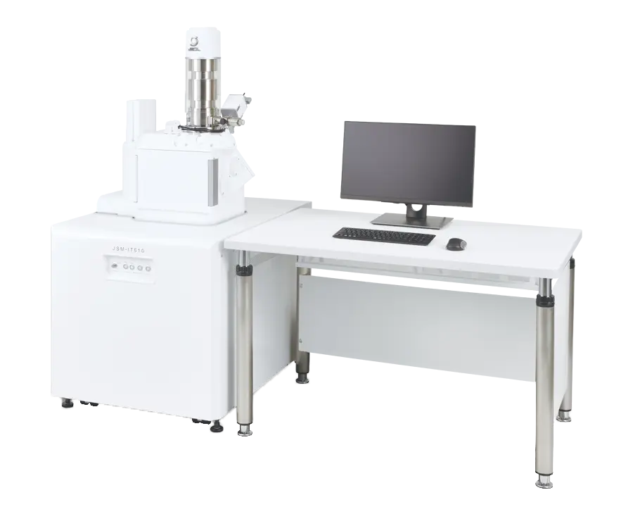
Manufacturer: JEOL — Japan
Overview
Scanning electron microscopes are indispensable tools not only for research but also for quality assurance and manufacturing, where the same observation routines are repeated day after day.
The JSM-IT510 is built around exactly that reality: its Simple SEM function lets the operator leave the repetitive manual steps of SEM observation to the instrument, making everyday imaging faster, easier and more consistent.
Features
Simple SEM
Just select the target field — Simple SEM supports daily routine work. The operator picks the field of view and the instrument performs the repetitive acquisition steps automatically, from a quick ×50 survey of an electronic device to a detailed ×1,000 image.
Specimen Exchange Navi
A guided workflow takes the operator from specimen exchange to automatic observation — safe and simple.
- Follow the Navi guide: The on-screen guide walks the operator through setting the specimen safely and correctly.
- Prepare during evacuation: Observation is prepared while the chamber evacuates. Options such as the Stage Navigation System (SNS), the large-sample SNSLS and the Chamber Scope (CS) extend the workflow.
- Observation starts automaticallyAfter evacuation the image is formed automatically — no manual start-up steps.
Zeromag
Zeromag simplifies navigation with a seamless transition from the optical image to the SEM image. The SEM image, optical image and holder graphic are all linked, giving a global view of every analysis location on the specimen. (The Stage Navigation System is required to display the optical image.).
Live Analysis / Live Map
Embedded EDS shows the spectrum or element maps in real time during image observation. The function supports searching for target elements and can raise an alert when they appear. Live Analysis is standard on the A (Analysis) and LA (Low Vacuum & Analysis) configurations.
Simple analysis
EDS analysis can be started within three clicks.
Variety of Advanced Options
Low-vacuum Hybrid Secondary Electron Detector (LHSED)
This detector collects both electron and photon signals, producing images with high signal-to-noise ratio and enhanced topographic information under low vacuum. (Requires the LV or LA configuration.).
Live 3D
Images obtained by the quadrant backscattered-electron detector can be displayed as a live 3D image, clearly representing the shape of a specimen even when its topography is subtle.
Montage
Automates large-area image collection and stitches the fields into one composite image — for example a 13 × 13 field montage of a fossil specimen.
Signal depth display
Displays the approximate analysis depth in the specimen — very useful when planning elemental analysis.
Four Configurations
The JSM-IT510 series can be equipped in four (04) configurations:
- BU (Base Unit) for observation under high vacuum;
- A (Analysis) with EDS attached as standard;
- LV (Low Vacuum) for high- and low-vacuum operation with backscattered-electron detector included;
- LA (Low Vacuum & Analysis) combining low vacuum, BED and EDS.
Typical Applications
Metals
Channeling contrast reveals crystal orientation in steel cross-sections, and fracture surfaces tell the story of failure: fatigue striations, ductile dimples and intergranular 'rock candy' patterns are all clearly resolved.
Semiconductors & Electronics
Resist-pattern cross-sections can be imaged and line widths measured for process quality control, while low-vacuum operation allows direct inspection of printed circuit boards without pre-treatment.
Polymers & Soft Materials
Low vacuum permits direct observation of non-conductive specimens such as superabsorbent polymers and mask fabrics; simultaneous SE and BE imaging separates shape from compositional information.
Biology
Cells and microorganisms — erythrocytes, chlorella, pollen — can be observed after chemical fixation or freeze-drying preserves their shape.
Specifications
| Specifications | JSM-IT510 |
|---|---|
| Resolution | High vacuum mode: 3.0 nm Low vacuum mode: 4.0 nm (BED) |
| Magnification | ×5 to ×300,000 |
| Electron gun | W filament, fully automatic gun alignment |
| Accelerating voltage | 0.3 kV to 30 kV |
| Objective lens aperture | Four stage, with XY fine adjustment function |
| Automatic function | Filament adjustment, Gun alignment adjustment Beam alignment Focus / Astigmatism / Brightness / Contrast correction |
| Energy dispersive X-ray spectrometer (EDS) (optional) | SD detector with detection surface area: 30/60/100 mm² can be installed |
| Major installable options | Backscattered Electron Detector (BED); Low Vacuum Hybrid Secondary Electron Detector (LHSED); Low Vacuum Secondary Electron Detector (LVSED); Energy dispersive X-ray spectrometer (EDS): SD detector with detection surface area: 30/60/100 mm²; Wavelength Dispersive X-ray Spectrometer (WDS); Electron Backscatter Diffraction (EBSD); Load Lock Chamber (pre-evacuation chamber); Stage Navigation System (SNS); Chamber Scope (CS); Operation Panel (OP2); LaB6 Electron Gun (LAB6); 3D Analysis Software (SVM) |