
120 kV TEM — Model: JEM-120i
The JEM-120i is a compact transmission electron microscope with simple operation — a useful tool for every user. It is designed around three ideas: compact, easy to use, and expandable.
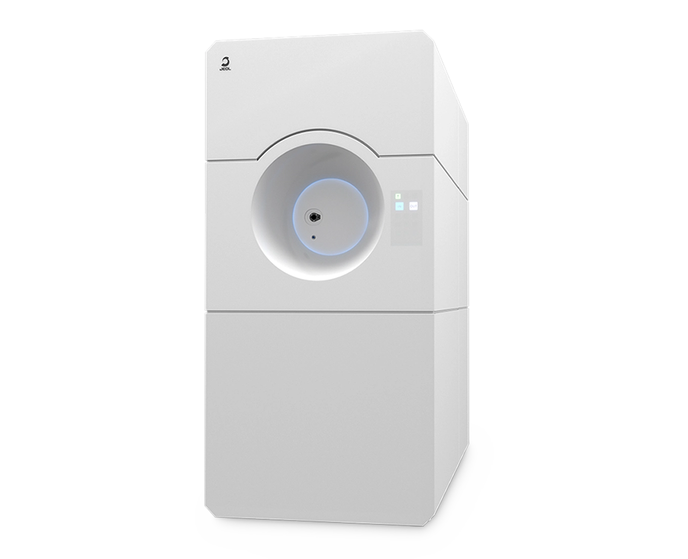
Manufacturer: JEOL — Japan
Overview
The JEM-120i is a compact transmission electron microscope with simple operation — a useful tool for every user. It is designed around three ideas: compact, easy to use, and expandable.
A totally new external appearance and compact size fit any installation location and support users from normal use through maintenance, while a wide range of retrofits lets you add new functions after purchase.
Specimen Observation in 4 Simple Steps
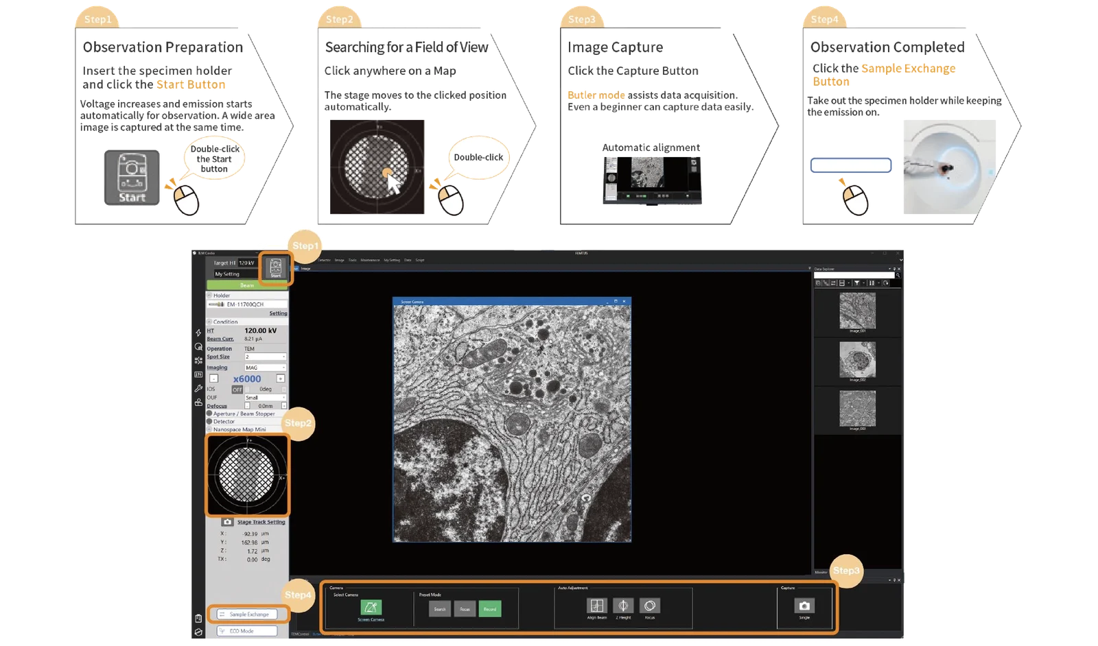
It takes only 4 steps from loading a specimen to completing observation.
Step 1 — Observation Preparation
Insert the specimen holder and click the Start button. Voltage increases and emission starts automatically for observation, and a wide-area image is captured at the same time.
Step 2 — Searching for a Field of View
Click anywhere on a Map and the stage moves to the clicked position automatically.
Step 3 — Image Capture
Click the Capture button. Butler mode assists data acquisition, so even a beginner can capture data easily, with automatic alignment.
Step 4 — Observation Completed
Click the Sample Exchange button and take out the specimen holder while keeping the emission on.
Easy To Use
Seamless Observation Without Switching Magnification Mode
The JEM-120i is equipped with an enhanced TEM control system and fully automated apertures, eliminating the need for switching magnification mode and selecting an aperture. Observation operations can be performed more smoothly than with previous models.
Drift Correction Function
In some cases a section shifts in one direction due to shrinkage or other reasons; capturing an image in this condition distorts it in the direction of the drift. The incorporated drift correction function helps users capture clear images — for example, of chitin nanofiber (negative staining) — and clearly resolves membrane structures such as the nuclear membrane and rough and smooth endoplasmic reticulum in rat liver (accelerating voltage: 120 kV).
"NeoView" Multi-Functional Digital Camera
The incorporated multi-functional digital camera "NeoView" enables observation with high sensitivity and low noise. A high frame rate and smooth field-of-view (FOV) search function let you find an FOV and acquire data without switching cameras — one camera from field-of-view search to data acquisition. It is also useful for specimen screening for cryo-observation.
Compact and User-Friendly
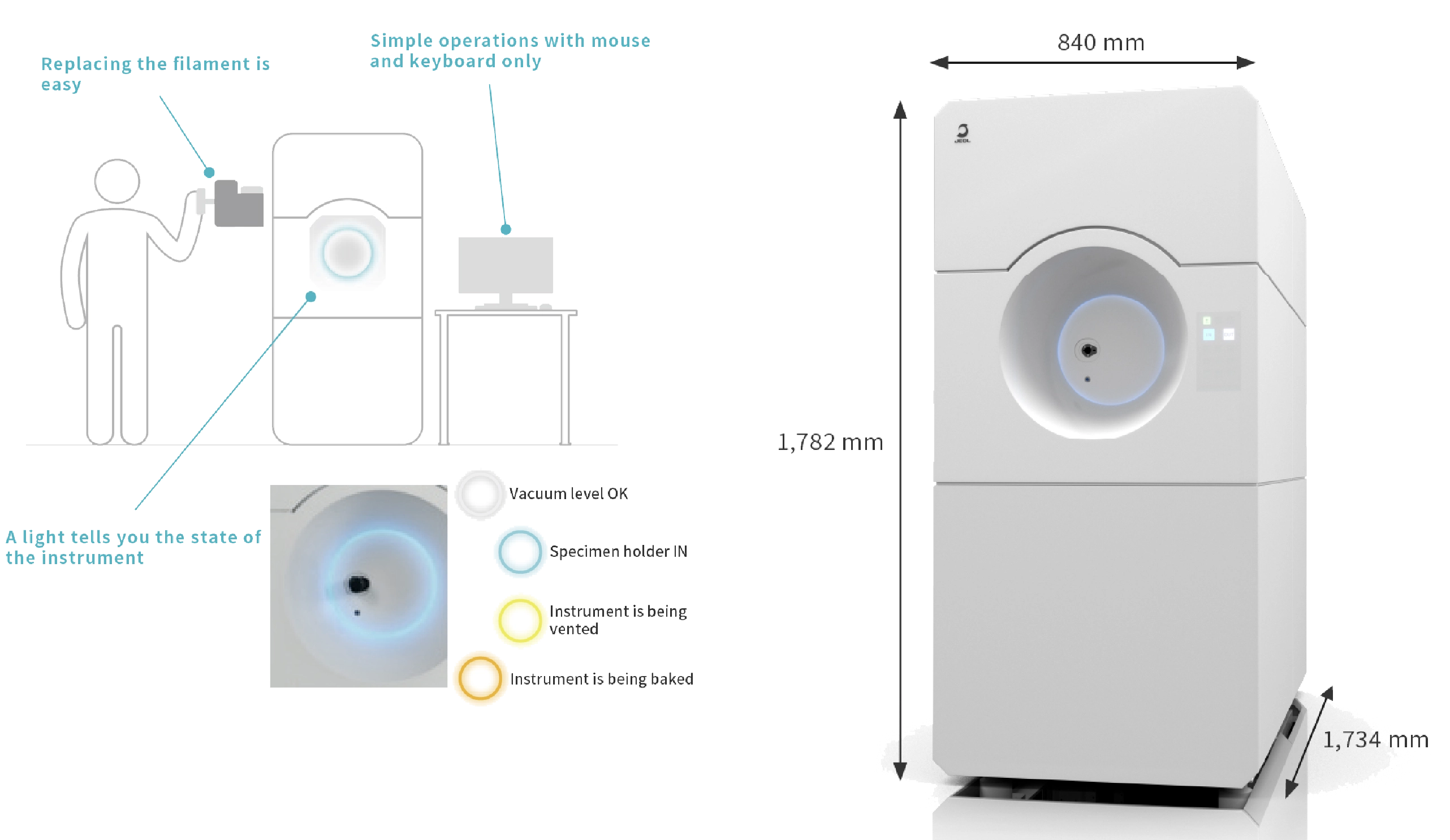
The size is more compact and operation has become more user-friendly, supporting users from normal use to maintenance.
Easy Filament Replacement
A newly developed cartridge-type filament unit makes filament replacement easy and safe. An auto conditioning function increases the accelerating voltage automatically after filament replacement.
Status Indicator Light
The instrument has an LED light on its front whose color changes depending on the state of the TEM — indicating states such as vacuum level OK, specimen holder in, instrument being vented, or instrument being baked — so you can determine its operating state at a distance.
Simple Mouse and Keyboard Operation
The new TEM control software allows the user to operate the TEM easily with a mouse and keyboard only. This eliminates the need for a dedicated operation panel* and is suitable for remote operation. *Operation panel can be configured as an option.
Energy Saving: ECO Mode
By switching to ECO mode when the instrument is not used, you can save power consumption by up to 80%* compared with when the TEM is in operation. *When accelerating voltage is 120 kV in TEM mode.
Expandable — A Variety of Options
The JEM-120i supports a wide range of retrofits and meets customers' needs of adding new functions after purchase.
SiN Window Chip Specimen Plate
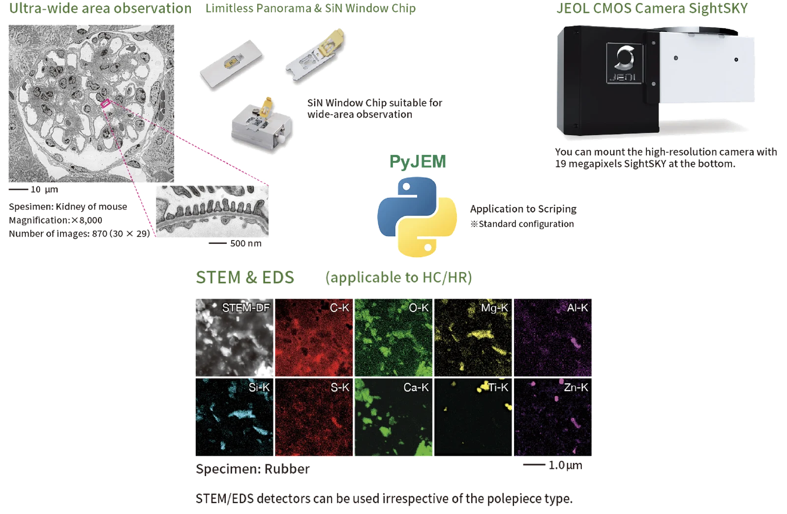
A specimen plate with supporting film for large-area observation, using high-strength SiN film. It has no grid bar and enables wide field-of-view observation at millimeter sizes. The stub is chemically resistant and the specimen can be stained in place.
SightSKY Fiber-Coupling CMOS Camera
JEOL's unique fiber-coupling CMOS camera can be mounted in addition to the incorporated NeoView camera. This high-resolution 19-megapixel camera produces high-definition images. Frame rate: 58 fps (all pixels read). Effective pixels: 19 M pixels (5,688 × 3,336 pixels). Image saving format: TIFF, BMP, JPG. Pixel size: 6.4 × 6.4 µm². Effective element size: 36.40 × 21.35 mm². Bottom cameras manufactured by other companies can be used — contact your JEOL service office for details.
Limitless Panorama (LLP) Ultra-Wide Field of View Montage
Limitless Panorama (LLP), a combination of beam scan and stage scan functions, enables automatic capture of images over an ultra-wide area with no limit — valuable for studying biological specimens whose complex structure requires both fine-structure and whole-tissue morphology. A link function with optical microscope images (LLP-CLEM) provides Correlative Light Electron Microscopy: positional alignment by selecting the same three or more points on both images. (Shot Meister manufactured by SIF is required separately.).
TEM Tomography (3D Structure Observation)
Captures a series of tilt images to acquire information about the internal structure of specimens three-dimensionally. Recorder acquires the continuous-rotation images required for 3D reconstruction of TEM images; Composer performs the 3D reconstruction; and Visualizer-evo visualizes the 3D data quickly and with high quality, even without a special rendering board. (Option: TEMography Function).
Specimen Holders
A lineup of holders can be selected according to your purpose: Quick Change Holder, Common Specimen Holder & Beryllium Analytical Retainer, Specimen Quartet Holder, High Tilting Specimen Trio Holder, High Tilting Holder, and Specimen Tilting Holder.
Two Versions
The JEM-120i offers two (02) versions:
- JEM-120i(HR) - is the high resolution version.
- JEM-120i(HC) - is the high contrast version.
Typical Applications
Life Science and Biology
Clear observation of membrane structures in rat liver, the basement membrane microstructure in mouse kidney (accelerating voltage 80 kV), thylakoid membrane and granum in carrot leaf chloroplasts, iron-containing ferritin protein particles, and ultra-wide-area montage of mouse spleen (25 × 25 images, ×8,000) and HeLa cells.
Materials Science
High-contrast observation of the sea-island structure of butadiene rubber in ABS resin (accelerating voltage 120 kV), chitin nanofiber, and 3D reconstruction of cylinder structures (en bloc staining in osmium tetroxide, tilt angle -70° to +70° in 1° steps).
Cryo Screening
The NeoView camera supports specimen screening for cryo-observation, including ice-embedded liposomes and negatively stained protein particles.
Specifications
| Specifications | JEM-120i(HC) | JEM-120i(HR) |
|---|---|---|
| Resolution | Point image: 0.38 nm Lattice image: 0.2 nm | Point image: 0.33 nm Lattice image: 0.14 nm |
| Accelerating voltage | Variable range from 20 to 120 kV with minimum step: 2.5 V | |
| Magnification: | ×50 to 1,200,000 | ×50 to 1,500,000 |
| Electron gun | Filament type: W Filament Unit/ LAB6 Filament Unit | |
| Power stability | Accelerating voltage: 2 × 10⁻⁶/min Objective lens: 1 × 10⁻⁶/min | |
| Minimum spot size | 0.2 μm in diameter |